Magnetic Resonance Imaging (MRI) is a powerful medical imaging technique that has revolutionized the field of healthcare. It allows clinicians to obtain highly detailed images of the internal structures of the human body, aiding in the diagnosis, monitoring, and treatment of various medical conditions. But how exactly does an MRI work? Let’s embark on a journey through the science behind this remarkable technology.
The Fundamentals: Magnetic Resonance Imaging
At its core, MRI relies on the principles of nuclear magnetic resonance (NMR), a phenomenon that occurs when certain atomic nuclei interact with a magnetic field. The nuclei in question, primarily hydrogen nuclei, have an inherent property called spin. When placed within a magnetic field, these nuclei align themselves with the field.
Step 1: Alignment with the Magnetic Field
The first step in an MRI involves aligning the hydrogen nuclei within the body’s tissues with the strong magnetic field created by the MRI machine. This alignment is the foundation of the entire imaging process. When the hydrogen nuclei align parallel to the magnetic field, they are in a lower energy state.
Step 2: Application of Radiofrequency (RF) Pulse
To capture images, the next step is to introduce radiofrequency (RF) energy in the form of a pulse perpendicular to the magnetic field. This RF pulse temporarily disrupts the alignment of the hydrogen nuclei. As a result, these nuclei absorb the RF energy, moving into a higher energy state.
Step 3: Relaxation: The Key to Imaging
Once the RF pulse is turned off, the hydrogen nuclei start returning to their low-energy state. This process, known as relaxation, involves two key components:
- T1 (Spin-Lattice) Relaxation: During T1 relaxation, the hydrogen nuclei transfer their absorbed energy to their surrounding environment. This causes them to realign with the magnetic field. T1 relaxation time varies among different tissues and is crucial for contrast in MRI images.
- T2 (Spin-Spin) Relaxation: T2 relaxation is another process through which the hydrogen nuclei release energy. The time it takes for these nuclei to return to their low-energy state after the RF pulse is turned off is called T2 relaxation time. Like T1, T2 relaxation times also vary between tissues and contribute to image contrast.
Step 4: The Resonance Signal
As the hydrogen nuclei return to their low-energy state, they emit the energy they absorbed in the form of radiofrequency signals. These signals are detected by the MRI machine’s receiver coils. The timing and strength of the signals are unique to each tissue type, forming the basis for image contrast.
Creating the Image: Spatial Encoding
To generate detailed images, MRI machines employ a technique called spatial encoding. This involves applying gradients of magnetic fields along different axes (X, Y, and Z) in a controlled manner. These gradients help determine the origin of each signal, enabling the MRI machine to create a 3D representation of the imaged area.
Image Reconstruction: Transforming Data into Pictures
The collected data is transformed into images through a complex process known as Fourier transformation. This mathematical technique is applied to the raw signals, converting them into grayscale images that reveal the structural and functional details of the imaged area.
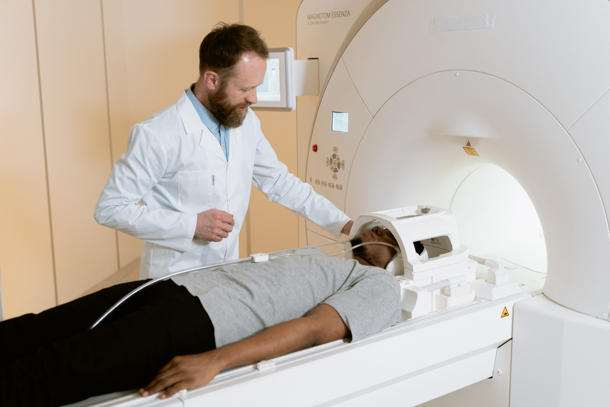
Key Components of an MRI Machine
An MRI machine comprises several vital components, including:
- Magnet: The large, powerful magnet generates the static magnetic field that aligns the hydrogen nuclei in the body.
- RF Coils: RF coils are used to transmit the radiofrequency pulse and receive the emitted signals.
- Gradient Coils: Gradient coils are responsible for creating the spatial encoding magnetic fields.
- Computer System: The computer system controls the entire MRI process, from pulse sequences to image reconstruction.
The Versatility of MRI
One of the most remarkable aspects of MRI is its versatility. By altering pulse sequences, parameters, and gradient fields, MRI can capture detailed images of various anatomical structures and physiological processes. From brain scans to musculoskeletal imaging, MRI has become an invaluable tool for healthcare professionals.
In conclusion, Magnetic Resonance Imaging is a marvel of modern medical technology. It relies on the inherent properties of hydrogen nuclei and their interaction with magnetic fields to produce high-resolution images of the human body. Understanding the fundamental principles behind MRI is the first step in appreciating its significant role in healthcare, aiding in accurate diagnoses and improved patient care.